branch

Branch Institutions

Topgene Research Institute

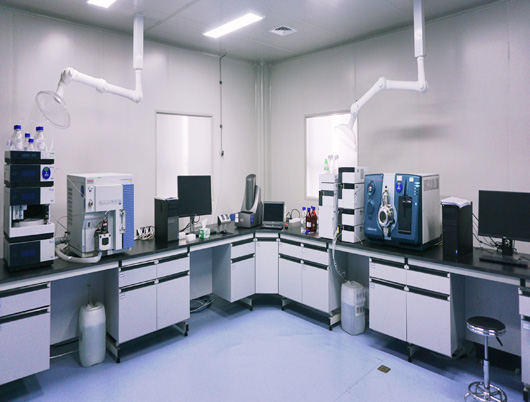
Hubei Topgene Biotechnology Research Institute Co., Ltd. (hereinafter referred to as Topgene Research Institute) is the core research and development institution of Hubei Topgene Biotechnology Co., Ltd. It is located at No. 12 Sandao Street, Wuchang District, Wuhan City (formerly within Hubei Pharmaceutical Industry Research Institute). With the core mission of "Service for Medical Innovation & Dedication to Life and Health", Topgene Research Institute focuses on the development of non-human primate (NHP) and rodent models for human diseases, providing services for the pharmacodynamics (PD), pharmacokinetics (PK), and pre-toxicity evaluation of various drugs (biotechnological drugs, chemical drugs, traditional Chinese medicine). It offers professional and efficient non-clinical drug research services for pharmaceutical companies, research institutes, and universities engaged in drug development. The institute covers approximately 2,800, including 1,000of  laboratories and 1,500 of animal facilities (280for barrier environments and 1,220for  conventional environments). The institute is capable of conducting experiments on conventional monkeys, dogs, small pigs, rabbits, as well as SPF-grade rats, mice, and guinea pigs. Topgene Research Institute currently has platforms for non-human primate surgery, behavioral studies and analysis, experimental animal molecular imaging, in vivo electrophysiology (under construction), cell molecular biology, biological analysis, and pathological analysis.


Service Platform

  • Monkey Surgery-related Platform

    1.Anaesthesia

    2.Multiple brain surgeries and brain stereotaxic infusion

    3.Lateral ventricle capsule implantation

    4.Splenectomy

    5.Ultrasonic-guided surgical technique

    6.Customized surgical services

    7.Intensive care


  • Monkey Behavioral Platform

    1.Cognitive ability measurement: WGTA (Wisconsin general test apparatus), Cantab (Cambridge neuropsychological test automated battery).

    2.Motor-related skill detection: Kurlan score, primate scan, fine finger coordination assay, food-grasping task.

    3.Emotion-related behavioral analysis: Spontaneous activity analyses, huddle, day-time sleeping, human intruder test, pace, turn in circles, face the wall, etc.


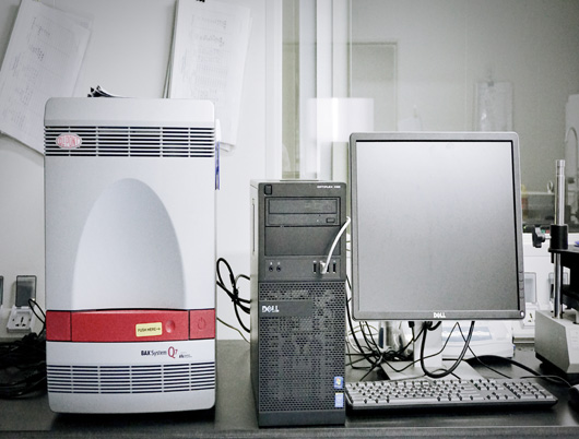
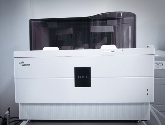
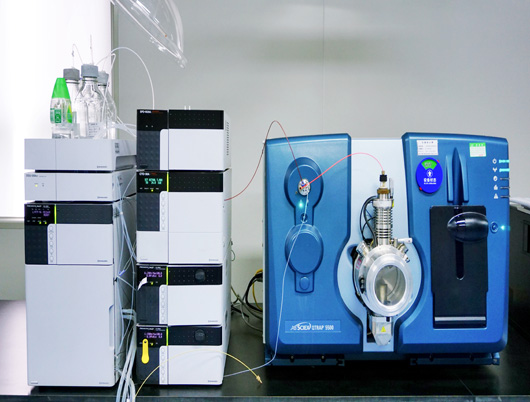
  • Bioanalysis Platform

    1.Antibody drug analyses

    2.Nucleic acid drug analyses

    3.Peptide drug analyses

    4.Biomarker analyses

    5.Anti-drug antibody analyses


  • Molecular Imaging Platform

    1.Radionuclide labeling and probe synthesis: 124I89Zr64Cu68Ga18F、AV45)、T807Tau.

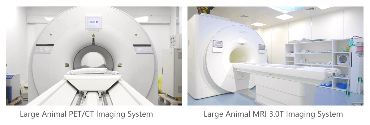
    2.Image acquisition, fusion and data analysis: PET/CT, MRI/fMRI, PET/MRI.

    3.Scope of application: PK/PD, ADME, dose range test and safety evaluation; disease model evaluation, pathogenesis study, drug targeting evaluation, etc.


Research Case

  • Macaca fascicularis model of Alzheimer's disease (AD)

    1. The increased level in Aβ42,Aβ42/Aβ40 and neurofilament light (NfL) in cerebrospinal fluid (CSF)the plasma detected by ELISA and MSD.

    2. The β-sheets enriched in the prefrontal cortex imaged by ThioS staining.

    3. The Aβ positive staining in prefrontal cortex (PFC) detected by 4G8 and 6E10 immunohistochemical staining.

    4. Astrocytes and microglia activation in the prefrontal cortex detected by immunohistochemistry.

    5. The increased level of phosphorylated Tau protein in the frontal lobe and hippocampus extracts detected by ELISA, Western blotting and immunohistochemistry.

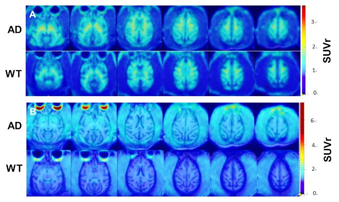
    6. Accumulation of Amyloid beta (Ab) and Tau in in the brain detected positron emission tomographyPETand magnetic resonance imaging (MRI).

    The increased Aβ (A) and Tau (B) accumulation in the model monkey brain detected by PET/MR imaging.

  • Rhesus monkey model of Parkinson's disease (PD)

    1. The PD-like behavioral phenotype evaluated by Kurlan score and nine-hole feeding tests.

    2. Effective attenuation of the PD-like phenotypes including Kurlan score and nine-hole feeding behaviors by application of L-DOPA.

  • Rhesus monkey model of major depressive disorder (MDD)

    1. The significantly increased huddling duration and stereotypical behaviors appeared respectively at 4 and 8 weeks after chronic mild unpredictable stress (CUMS) modeling.

    2. The significantly increased plasma levels of IL-6 and TNF-α detected at 10 weeks after chronic mild unpredictable stress (CUMS) modeling. 

  • Cynomolgus monkey of metabolic dysfunction-associated steatotic liver disease (MASLD)

    1. The HE staining of liver tissue showed fatty degeneration at 3 months after modeling.


                                    Control                                                 Model

          Figure 1. The HE staining of liver tissue in cynomolgus monkey with non-alcoholic Fatty Liver


    2. The HE staining showed fat vacuolization and extensive infiltration of inflammatory cells at 5-6 months after modeling.


                                    Control                                                  Model

          Figure2.The HE staining of liver tissue in cynomolgus monkey with non-alcoholic Steatohepatitis.


    3. The VG staining of liver tissue showed destruction of lobular structure and formation of fibrous septa at 8-9 months after modeling.


         Figure 3. The VG staining of liver tissue in cynomolgus monkey with non-alcoholic liver fibrosis.

  • Cynomolgus monkey of hemophilia

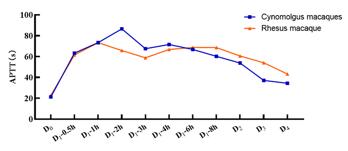
    1. The significantly prolonged APTT time up to four times longer than before modeling.

                                                      Figure 1. Change in APTT after modeling.


            2. The significantly increased area of subcutaneous bruising after modeling.


                                                      Figure 2. Change in bruised area after modeling.


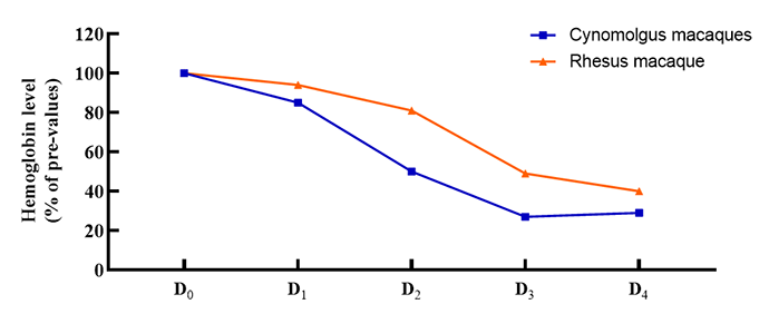
            3.The significantly reduced hemoglobin levels after modeling.


                                                      Figure 3. Change of hemoglobin level after modeling.


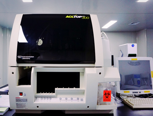
Infrastructure and Equipment