


 Service Capabilities
- 10/23/2023 12:24:35 PM | Visits:1145
Macaca fascicularis model of Alzheimer's disease (AD)
In addition to learning and memory deficits, our AD NHP model started to show elevated levels of Aβ42, Aβ42/40 and NfL at 1 month after modeling in cerebrospinal fluid samples measured by conventional ELISA or/and MSD. The elevations of Aβ42, Aβ42/40 and NfL were also detected at 6 months after modelling, and the increases were still detectable at 12 months after the modeling. We also detected Aβ and Tau accumulations in prefrontal cortex and the hippocampus by PET scan at 12 months after modelling.
Compared to other reported NHP AD models in literatures, the main advantages of our AD models are as follows:
● Our AD models show simultaneously abnormal ATN biomarkers (i.e., elevated Abeta, pTau, and NFL both in CSF and the periphery blood) and in the brain, and as well as behavioral (learning and memory) deficits, which perfectly fulfill the clinical diagnostic standards of AD patients.
● Our AD models can be replicated in large quantities with reasonable reproducibility to fulfill the CRO services, which is currently almost not available by transgenic techniques in monkeys.
● By the aid of MRI-PET imaging technique, we can provide consecutive data to show dynamic drug effects with no need to sacrificing the precious and expensive monkeys.
● We can design and provide custom-made AD models according to the customers’ requirements.
● The procures we developed for producing AD monkey models show high success rate with low mortality, and the memory deficits and brain pathologies can sustain for a long period of time, which provides guarantee for evaluating the efficacy of investigational drugs.
1. The increased level in Aβ42,Aβ42/Aβ40 and neurofilament light (NfL) in cerebrospinal fluid (CSF) and the plasma detected by ELISA and MSD.
2. The β-sheets enriched in the prefrontal cortex imaged by ThioS staining.
3. The Aβ positive staining in prefrontal cortex (PFC) detected by 4G8 and 6E10 immunohistochemical staining.
4. Astrocytes and microglia activation in the prefrontal cortex detected by immunohistochemistry.
5. The increased level of phosphorylated Tau protein in the frontal lobe and hippocampus extracts detected by ELISA, Western blotting and immunohistochemistry.
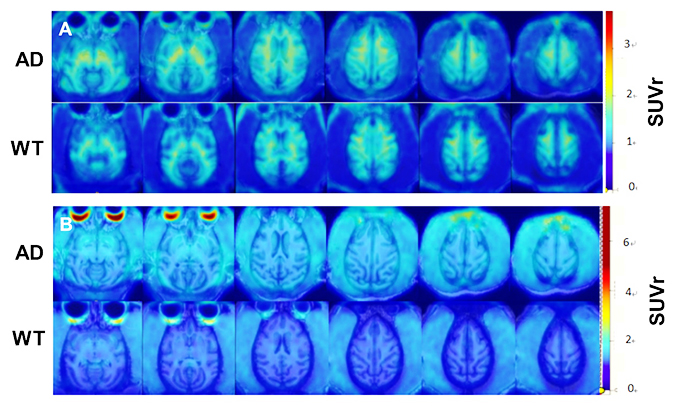
6.The increased Aβ (A) and Tau (B) accumulation in the model monkey brain detected by PET/MR imaging.
- Previous article:None
- Next article:Rhesus monkey model of Parkinson's disease (PD)