


 Service Capabilities
- 4/30/2024 12:48:11 PM | Visits:990
Cynomolgus monkey of metabolic dysfunction-associated steatotic liver disease (MASLD)
Nonalcoholic fatty liver, as the manifestation of metabolic syndrome in liver, is closely related to obesity, insulin resistance (IR), diabetes, hyperlipidemia and other metabolic abnormalities. NAFLD is a clinicopathologic syndrome characterized by hepatic parenchymal cell steatosis and fat storage. The disease spectrum includes simple hepatic steatohepatitis, non-alcoholic steatohepatitis (NASH), liver fibrosis, which can progress to cirrhosis and liver cancer.
Molding methods: High fat diet was given and CCl4 was injected subcutaneously to induce liver fibrosis and fatty degeneration.
Detection index:
Weekly measurement: weight, food intake;
Regular tests:liver function, liver B-ultrasound, liver puncture pathology.
1. The HE staining of liver tissue showed fatty degeneration at 3 months after modeling.
Control Model
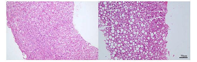
Figure 1. The HE staining of liver tissue in cynomolgus monkey with non-acoholic Fatty Liver.
2. The HE staining showed fat vacuolization and extensive infiltration of inflammatory cells at 5-6 months after modeling.
Control Model
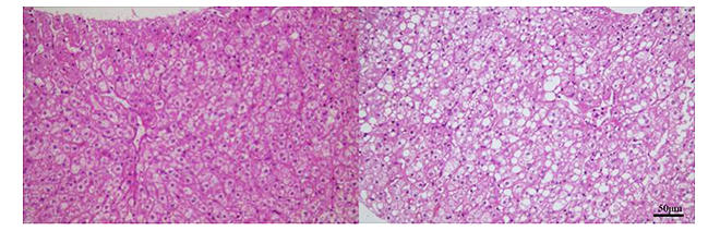
Figure 2. The HE staining of liver tissue in cynomolgus monkey with non-alcoholic Steatohepatitis.
3. The VG staining of liver tissue showed destruction of lobular structure and formation of fibrous septa at 8-9 months after modeling.
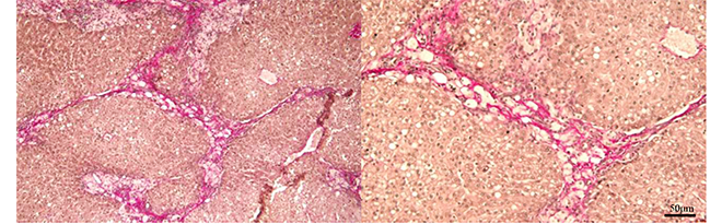
Figure 3. The VG staining of liver tissue in cynomolgus monkey with non-alcoholic liver fibrosis.
- Previous article:Cynomolgus monkey of hemophilia
- Next article:Rhesus monkey model of major depressive disorder (MDD)